The Spatial Mind · Part 4
Your Mind Is a Map: The Positioning System Inside Your Head
Recordings in rats revealed cells sensitive to place, direction, and distance. These findings help explain spatial navigation and offer clues about memory, with limits that matter.
The question: What neural evidence connects memory to navigation?
Big idea: Place, grid, direction, boundary, speed, and time signals support navigation; hippocampal memory is closely tied to this mapping machinery.
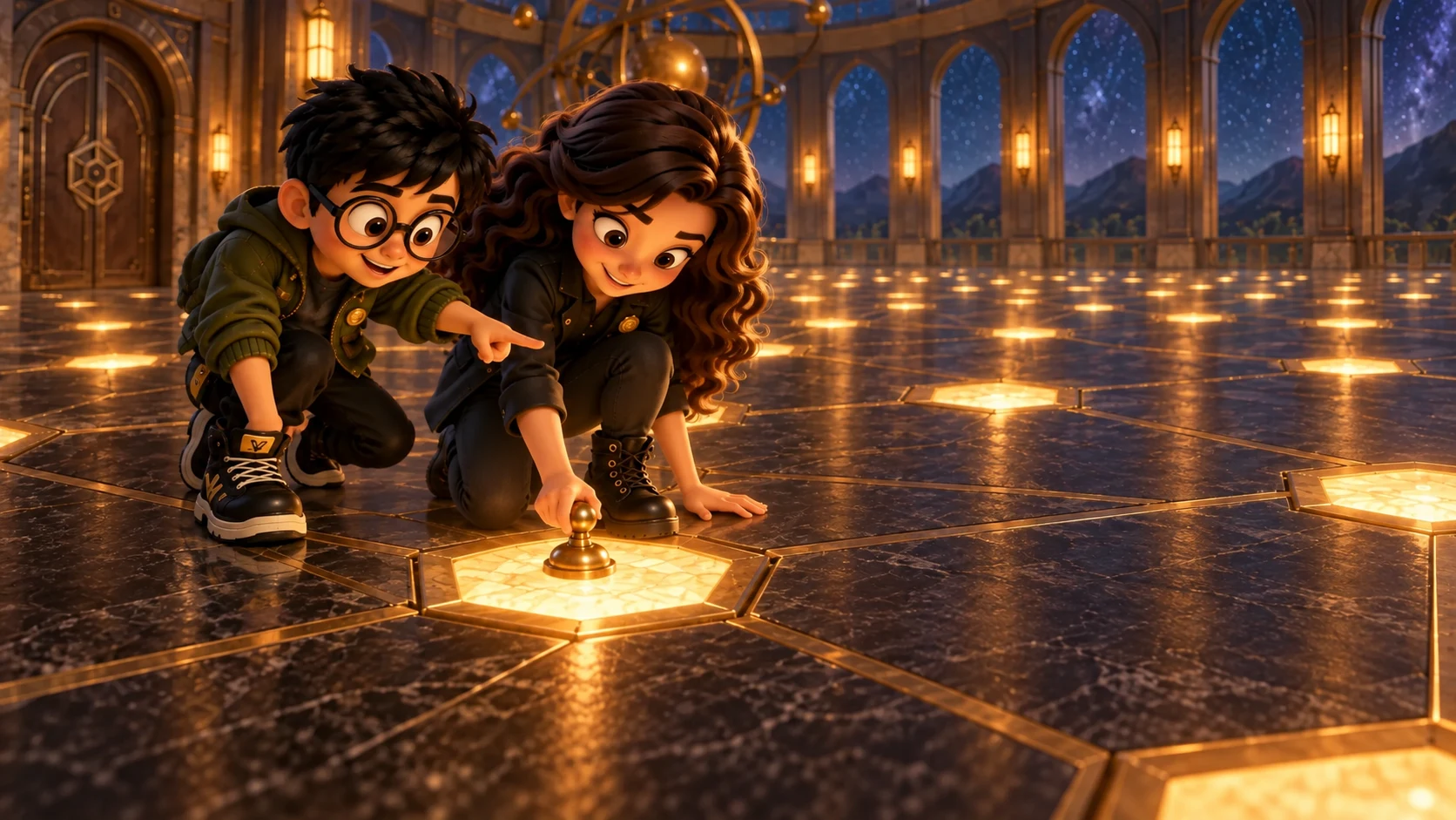
In 1948, Edward Tolman published a paper with the wonderful title Cognitive Maps in Rats and Men.
Many psychologists of the time explained learning through stimulus, response, and reward. Tolman pointed to maze experiments by Blodgett and by Tolman and Honzik: rats that had explored without food made many errors, then their error counts dropped sharply after food appeared. The result suggested that the animals had learned something during unrewarded exploration that their earlier performance did not reveal.
Tolman proposed a cognitive map: knowledge of relationships in the environment, broader than a memorized sequence of turns. His paper also described route-blocking experiments in which rats often chose a path pointing toward the goal's former location, though their choices were not always precise.
That was a behavioral interpretation, not a recording of a map. Twenty-three years later, neural recordings offered a new kind of evidence about how location can be represented.
A cell with a place field
In 1971, John O'Keefe and Jonathan Dostrovsky were recording from single neurons in the hippocampus of a freely moving rat. O'Keefe later described the moment of realising that the cells "weren't particularly interested in what the animal was doing or why it was doing it but rather... where it was in the environment."
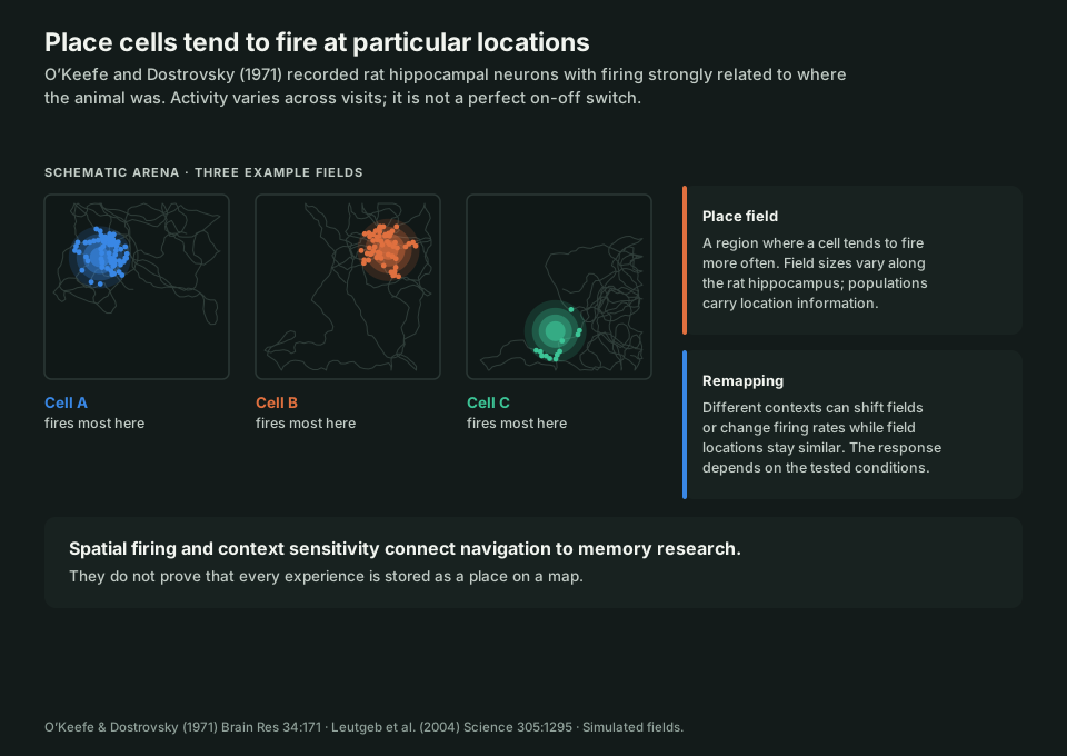
Some recorded cells fired much more when the rat passed through a particular region than elsewhere in the chamber. The firing was location-sensitive, though not an on-off switch that behaved identically on every visit.
They are called place cells, and the region where a cell tends to fire is its place field. Across a population, their activity carries information about the animal's location in a studied environment.
Two details that matter for the rest of this series.
Field size is not fixed. In rats, field size varies along the hippocampus. On an 18-metre track, Kjelstrup and colleagues measured fields from under a metre dorsally to much larger fields ventrally. Different scales can represent location with different spatial detail.
Activity can change with context. In some environment changes, place fields shift or appear and disappear, a pattern called global remapping. In others, firing rates change while field locations remain similar, called rate remapping. These are observed patterns, not a fixed rule that every new room or wall colour triggers one particular response.
Those findings connect location to context, one reason the hippocampus matters to both navigation and memory. A place cell's firing alone does not tell us how a whole experience is stored.
The hexagons
Place-cell activity raises a question: what other signals help an animal track its location? Self-motion and landmarks both matter, and nearby entorhinal regions contain another striking spatial pattern.
In 2005, Torkel Hafting, Marianne Fyhn, Sturla Molden, May-Britt Moser, and Edvard Moser recorded from medial entorhinal cortex and described grid cells.
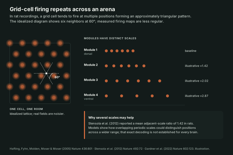
A single cell fired at multiple locations that formed an approximately regular triangular pattern across the tested arena. Idealized diagrams show six neighboring fields at 60-degree intervals; actual firing maps are noisier.
Their repeated firing pattern is one component of the brain's spatial representation.
Grid cells are organized in modules with similar spacing and orientation. In rats, Stensola and colleagues reported discrete scales with a mean ratio of 1.42 between adjacent modules, close to √2. Combining several periodic scales is a proposed way to represent location over a larger range; the measured ratio does not prove a single exact coordinate algorithm for every brain.
The rest of the committee
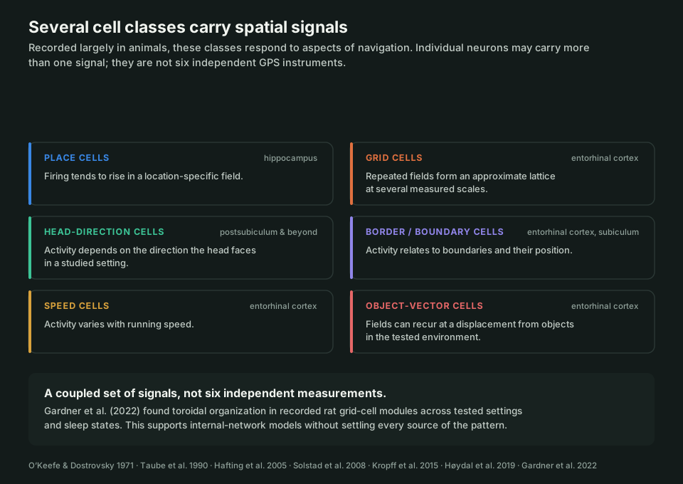
"Inner GPS" is the Nobel Committee's phrase, from the 2014 press release. It is a useful hook, though the cells below were identified largely through animal recordings and do not behave as six separate instruments with one job each:
- Place cells (hippocampus) show location-sensitive firing.
- Grid cells (entorhinal cortex) show repeated spatial firing fields at several scales.
- Head-direction cells (including postsubiculum) are sensitive to the direction the head faces.
- Border and boundary-vector cells respond to environmental boundaries or their relative position.
- Speed cells have activity related to the animal's running speed.
- Object-vector cells fire at particular displacements from objects in tested environments.
The 2014 Nobel Prize in Physiology or Medicine went to O'Keefe and the Mosers "for their discoveries of cells that constitute a positioning system in the brain."
In 2022, Gardner and colleagues recorded hundreds of grid cells in rats and analyzed the population activity. Individual recorded modules showed a toroidal organization, with related structure across the environments and sleep states they tested. This supports models with internally organized network dynamics; it does not settle every source of the grid pattern or show that human thought is arranged on a torus.
The taxi drivers, told properly
You have heard about this study. You have probably heard it wrong.
London cab drivers spend three to four years memorising 25,000 streets to pass a test called the Knowledge. In 2000, Eleanor Maguire's group compared 16 of them to controls.
The finding was regional rather than an overall "bigger hippocampus": posterior hippocampus was larger and anterior smaller in that sample. Right posterior volume correlated positively with months of driving (r = 0.6); anterior volume correlated negatively (r = −0.6). This cross-sectional comparison could not establish what changed within individuals.
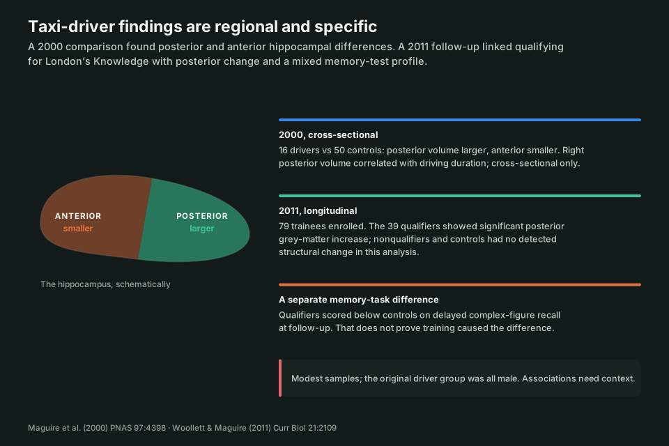
The 2011 study adds longitudinal evidence. Woollett and Maguire followed 79 trainees and 31 controls; 39 trainees qualified, 20 did not, and the others did not complete the follow-up. The qualifiers showed a significant posterior grey-matter increase, while the nonqualifiers and controls did not show a detected structural change. Because people were not randomly assigned to qualify, this strengthens a learning-related interpretation without isolating every cause.
The qualification group also scored below controls on delayed recall of a complex figure at follow-up. That is a difference on one visual memory task, not proof that navigation training damaged their memory.
The studies were modest in size, and the original driver group was all male. Together they show a more specific picture than a general brain-growth headline: spatial expertise was associated with regional hippocampal differences and a mixed memory-test profile.
Time, as well as space
One more piece, and it is the hinge into Part 5.
The hippocampus does not only respond to position. In 2008, Pastalkova and colleagues recorded sequences of hippocampal activity while rats ran on a wheel during a delay. In 2011, MacDonald and colleagues reported time cells that were active at successive moments within a task delay. Their activity also related to location and behavior, so the result is richer than a pure stopwatch signal.
Together, the findings show that hippocampal activity can carry spatial and temporal information in these tasks. They help explain why the hippocampus matters to remembering experiences, but they do not reduce an episode to two cell types or establish one indexing mechanism for all memory.
Which raises the question Part 5 is about.
If navigation-related circuits can represent physical position and task sequence, can they also help represent learned relationships with no physical floor?
Part 5 tests that question using specific studies of conceptual, social, sound, and odour relationships.
Sources
- Tolman, E. C. (1948). Cognitive maps in rats and men. Psychological Review, 55(4), 189–208.
- O'Keefe, J., & Dostrovsky, J. (1971). The hippocampus as a spatial map. Brain Research, 34, 171–175.
- O'Keefe, J. (2014). Spatial cells in the hippocampal formation. Nobel Lecture.
- O'Keefe, J., & Nadel, L. (1978). The Hippocampus as a Cognitive Map.
- Leutgeb, S., et al. (2005). Independent codes for spatial and episodic memory in hippocampal neuronal ensembles. Science, 309(5734), 619–623.
- Hafting, T., Fyhn, M., Molden, S., Moser, M.-B., & Moser, E. I. (2005). Microstructure of a spatial map in the entorhinal cortex. Nature, 436, 801–806.
- Stensola, H., et al. (2012). The entorhinal grid map is discretized. Nature, 492, 72–78.
- Taube, J. S., Muller, R. U., & Ranck, J. B. (1990). Head-direction cells. J Neurosci, 10(2), 420–435.
- Solstad, T., et al. (2008). Representation of geometric borders in the entorhinal cortex. Science, 322, 1865–1868.
- Kropff, E., et al. (2015). Speed cells in the medial entorhinal cortex. Nature, 523, 419–424.
- Høydal, Ø. A., et al. (2019). Object-vector coding in the medial entorhinal cortex. Nature, 568, 400–404.
- Gardner, R. J., et al. (2022). Toroidal topology of population activity in grid cells. Nature, 602, 123–128.
- Maguire, E. A., et al. (2000). Navigation-related structural change in the hippocampi of taxi drivers. PNAS, 97(8), 4398–4403.
- Woollett, K., & Maguire, E. A. (2011). Acquiring "the Knowledge" of London's layout drives structural brain changes. Current Biology, 21(24), 2109–2114.
- Kjelstrup, K. B., et al. (2008). Finite scale of spatial representation in the hippocampus. Science, 321(5885), 140–143.
- Pastalkova, E., et al. (2008). Internally generated cell assembly sequences. Science, 321, 1322–1327.
- MacDonald, C. J., Lepage, K. Q., Eden, U. T., & Eichenbaum, H. (2011). Hippocampal "time cells" bridge the gap in memory for discontiguous events. Neuron, 71(4), 737–749.
- Eichenbaum, H. (2014). Time cells in the hippocampus. Nature Reviews Neuroscience, 15, 732–744.
- The Nobel Prize in Physiology or Medicine 2014. NobelPrize.org.